Evaluating NAD+ for Nonclinical Laboratory Research
Nicotinamide adenine dinucleotide in its oxidized form, NAD+, is a coenzyme in redox reactions and a substrate for enzymes including sirtuins and poly(ADP-ribose) polymerases. For laboratories studying nucleotide chemistry, enzyme activity, or cellular metabolism, reagent characterization is an important part of experimental design.
Sourcing NAD+ requires more than comparing advertised purity percentages. Identity, chromatographic purity, quantitative content, degradation products, and storage history answer different questions. A “research-grade” label alone does not define these properties or establish suitability for a particular assay. This guide focuses on documentation and analytical checks for nonclinical laboratory work, not on administration or clinical applications.
Chemical Identity and Degradation
NAD+ contains two nucleotide units joined through their phosphate groups: one contains adenine and the other nicotinamide. The commonly listed molecular formula is C21H27N7O14P2, with a molar mass of approximately 663.43 g/mol. Procurement and analytical records should specify the supplied chemical form, including any counterions or hydration, rather than assuming that every powder has the same composition per unit mass.
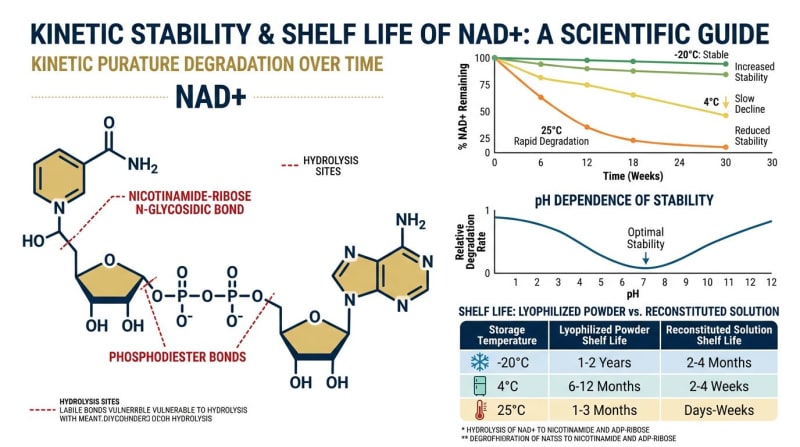
Cleavage of the nicotinamide–ribose bond can produce nicotinamide and ADP-ribose. Degradation therefore changes both the amount of intact NAD+ and the composition of the sample. In a cell-free enzyme assay, these changes can complicate interpretation of substrate-dependent activity. The relevance of any impurity must be evaluated for the specific enzyme, assay conditions, and endpoint rather than inferred from a total purity percentage.
Temperature, pH, moisture exposure, sample matrix, and elapsed time can influence stability. There is no universal temperature or pH cutoff that establishes stability for every formulation. Storage conditions and retest intervals should be supported by documentation for the supplied form and, where necessary, by laboratory stability checks.
HPLC: Assessing Separation and Chromatographic Purity
High-performance liquid chromatography separates sample components through their interactions with a stationary phase and mobile phase. For NAD+, the method should resolve the target from relevant impurities and degradation products. The presence of a large main peak does not, by itself, establish identity or quantify all material in the container.
When reviewing certificates of analysis, request the chromatogram and method information behind the reported result. Peak-area percentage is detector- and method-dependent: compounds may have different response factors, some components may not be detected, and unresolved impurities may contribute to the main peak. Chromatographic area percentage is not automatically equivalent to mass fraction or quantitative NAD+ content.
What to Review in a Chromatogram
- Peak assignment: Look for comparison with a characterized reference material and complementary identity data. Retention time alone is not definitive identification.
- Resolution: Check whether the method separates NAD+ from nicotinamide, ADP-ribose, and other relevant components. A clean-looking peak does not exclude co-elution.
- Acquisition and integration: Review detector settings, run duration, integration rules, and reporting thresholds. These affect the reported area percentage.
- Blanks and system suitability: Examine evidence for adequate repeatability, resolution, and control of carryover. A flat baseline does not prove the absence of contaminants.
- Acceptance criteria: Define criteria for the intended laboratory assay. Neither 98% nor 99% area purity is a universal threshold for experimental suitability.
| Parameter | Information to request | Interpretive limitation |
|---|---|---|
| Separation conditions | Column chemistry, mobile phases, gradient or isocratic conditions, and relevant resolution data | A C18 or HILIC designation alone does not establish adequate retention or separation. |
| Detection | Detector type and wavelength; UV detection near 260 nm is commonly useful for nucleotide analysis | UV absorbance is not unique to NAD+ and does not detect every impurity. |
| Purity result | Calculation basis, chromatograms, integration rules, and reporting limits | Area normalization does not establish total content or exclude undetected components. |
| Quantitative assay | Reference material, calibration, recovery, and measurement basis | Absolute content requires a suitable quantitative method, not just a normalized peak area. |
Mass Spectrometry: Supporting Identity
Mass spectrometry measures ion mass-to-charge ratios, not the bulk molecular weight directly. Interpretation of an electrospray spectrum requires the ionization polarity, charge state, adduct assignment, and expected ion composition. A spectral peak should not be rejected simply because its mass-to-charge ratio differs from the neutral-form molar mass.
LC-MS combines chromatographic separation with mass-spectral information. It can help distinguish NAD+ from related compounds such as NADH and NADP+ when separation, ion assignments, and reference data are appropriate. Accurate mass and fragmentation data can strengthen an identity assignment, but no single mass peak establishes complete purity or excludes every structural alternative.
Quantitative LC-MS additionally requires suitable calibration and assessment of matrix effects, recovery, and sample stability. Signal intensity alone is not a concentration measurement, and MS response differences can make uncalibrated comparisons misleading.
Distinguishing NAD+ from Related Research Materials
NAD+, NADH, NADP+, nicotinamide mononucleotide (NMN), and nicotinamide riboside (NR) are chemically distinct materials. They should not be treated as interchangeable reagents or selected on the assumption of equivalent biological effects. Purchasing records should identify the exact target compound and chemical form required by the experimental question.
For cell-free assays, reagent selection should follow the enzyme reaction and analytical endpoint being measured. For in-vitro cell experiments, extracellular addition alone does not establish intracellular exposure or compartment-specific concentrations. Those quantities require appropriate measurements and controls. Neither type of experiment establishes human or veterinary suitability.
A Batch-Level Procurement Checklist
A certificate of analysis is useful only to the extent that its methods, results, and sample traceability support the proposed experiment. Supplier reputation, a catalogue description, or a generic example report cannot substitute for documentation tied to the purchased lot.
- Identity and traceability: Match the container label, batch number, chemical form, and report. Check analysis dates and whether testing applies to the actual supplied lot.
- Quantitative content: Distinguish the amount of NAD+ from total powder mass. Water, counterions, and residual material can affect mass-based calculations.
- Relevant impurities: Request results for degradation products or process-related impurities that could interfere with the planned assay. The absence of a listed impurity is not necessarily evidence that it was tested.
- Residual solvents: Request an appropriate method and numerical results where solvents are relevant. A routine UV chromatogram is not a comprehensive residual-solvent assessment; “safe ppm levels” are not a universal laboratory specification.
- Appearance: Compare the material with its documented description. An unexpected change warrants investigation, but color or clarity alone cannot identify degradation or confirm purity.
- Microbial and endotoxin attributes: For in-vitro experiments sensitive to these variables, establish appropriate specifications and check whether testing was performed. Chemical purity does not establish sterility or low endotoxin content. Endotoxin results also require attention to assay interference and recovery.
- Stability and transport: Request storage conditions, packaging information, and the basis of any retest date or shelf-life claim. Investigate temperature excursions against available stability evidence rather than assuming an outcome.
The quality control information and available batch records can be starting points for document review. This guide does not establish that any particular Alpha Carbon Labs lot has undergone a specified test or meets an experimental acceptance criterion.
Laboratory Handling, Storage, and Hazard Review
Follow the applicable safety data sheet, institutional chemical-hygiene procedures, and documented storage instructions for the supplied material. Assess handling hazards before opening the container; use appropriate protective equipment and minimize powder dispersal and contact with skin or eyes. Incomplete hazard information should not be interpreted as evidence that a material is harmless.
Record receipt conditions, lot identifiers, opening dates, and storage history. Keep containers appropriately closed to limit contamination and uncontrolled moisture exposure. For laboratory samples, document time outside validated storage conditions and any repeated temperature changes. Use analytical reassessment when the available records cannot establish continued suitability.
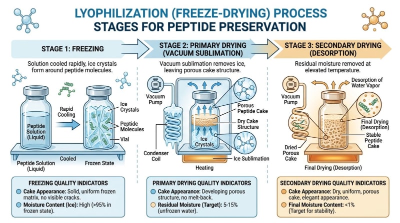
Do not assign a universal frozen-storage temperature or a months-to-years shelf life without supporting data. Likewise, lyophilization does not independently establish stability, sterility, or retained chemical content. Packaging integrity and residual moisture remain relevant variables.
Using Analytical Evidence Without Overinterpreting It
Identity testing, chromatographic purity, quantitative content, and stability testing provide complementary evidence. None alone guarantees an interference-free experiment. Reference materials, analytical blanks, relevant assay controls, and lot-to-lot comparisons help laboratories assess whether reagent variation affects their results.
Literature interpretation requires a separate check: findings from cell-free systems, cultured cells, animals, human study populations, and computational models must remain tied to their actual evidence source. Reviews summarize multiple studies and should not be treated as a single experimental model. The retained bibliography below does not constitute batch testing or establish properties of Alpha Carbon Labs materials.
Alpha Carbon Labs supplies materials solely for nonclinical laboratory research. Procurement decisions should be based on documented analytical attributes and the requirements of the laboratory method, without implying clinical approval or intended human or veterinary use.
References
- Yaku, K., Okabe, K., & Nakagawa, T. (2018). NAD metabolism: Implications in aging and longevity. Ageing Research Reviews, 47, 1-17. — https://doi.org/10.1016/j.arr.2018.05.003
- Trammell, S. A., & Brenner, C. (2013). Targeted, LCMS-based metabolomics for quantitative measurement of NAD+ metabolites. Computational and Structural Biotechnology Journal, 4, e201301012. — https://doi.org/10.5936/csbj.201301012
- Ratajczak, J., et al. (2016). NR and NMN: Do they increase NAD+ in mammalian cells? A systematic review. Metabolism, 65(12), 1774-1786. — https://doi.org/10.1016/j.metabol.2016.09.006
- Yoshino, J., Baur, J. A., & Imai, S. I. (2018). NAD+ intermediates: The biology and therapeutic potential of NMN and NR. Cell Metabolism, 27(3), 513-528. — https://doi.org/10.1016/j.cmet.2018.01.011
- Canto, C., & Auwerx, J. (2012). Targeting sirtuin 1 to improve metabolism: all you need is NAD+? Pharmacological Reviews, 64(1), 166-187. — https://doi.org/10.1124/pr.110.003905
- Sporty, J. L., et al. (2008). High-performance liquid chromatography separation of nicotinamide adenine dinucleotide (NAD+) and its distinct degradation products. Journal of Chromatography B, 874(1-2), 15-22. — https://doi.org/10.1016/j.jchromb.2008.08.025
- Navarro, F., et al. (2013). Stability of Nicotinamide Adenine Dinucleotide (NAD+) in solution under different conditions. International Journal of Pharmaceutics, 452(1), 265-271. — https://doi.org/10.1016/j.ijpharm.2013.04.072
- Covarrubias, A. J., Perrone, R., Grozio, A., & Verdin, E. (2021). NAD+ metabolism and its roles in cellular processes during ageing. Nature Reviews Molecular Cell Biology, 22(2), 119-141. — https://doi.org/10.1038/s41580-020-00313-x
- Szeto, H. H. (2014). First-in-class cardiolipin-protective compound as a therapeutic agent to restore mitochondrial bioenergetics. British Journal of Pharmacology, 171(8), 2029-2050. — https://doi.org/10.1111/bph.12461
- Lee, C., et al. (2015). The mitochondrial-derived peptide MOTS-c promotes metabolic homeostasis and reduces diet-induced obesity. Cell Metabolism, 21(3), 443-454. — https://doi.org/10.1016/j.cmet.2015.02.009
- Khavinson, V. K., et al. (2003). Epithalon peptide induces telomerase activity and telomere elongation in human somatic cells. Bulletin of Experimental Biology and Medicine, 135(6), 590-592. — https://doi.org/10.1023/a:1025493705728
Limitations
Published findings do not establish the safety, effectiveness, approval, or suitability of material sold by Alpha Carbon Labs for human or veterinary use. Laboratory suitability requires batch-specific evidence and method-specific evaluation.