Semaglutide vs Retatrutide for Adipose Tissue Research
Semaglutide and retatrutide differ in receptor target profile, making them distinct subjects for nonclinical investigations of receptor signaling and adipose biology. A useful comparison separates receptor activity from tissue-level observations and distinguishes direct cellular responses from changes occurring across an intact organism.
Receptor Identity and Research Scope
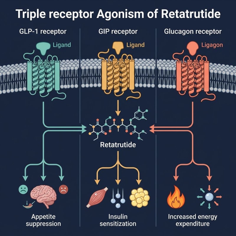
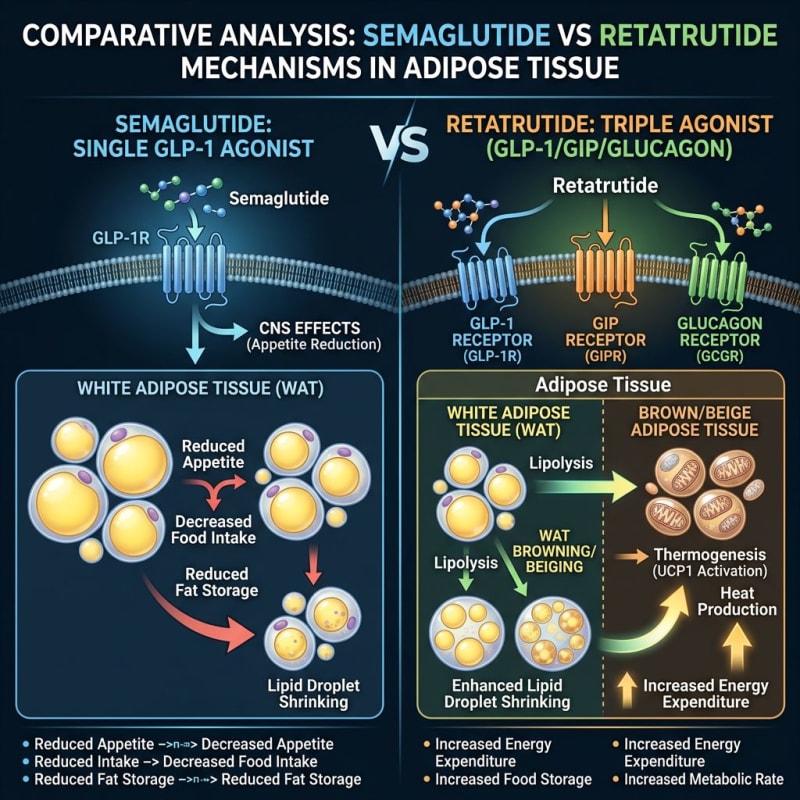
Semaglutide is a glucagon-like peptide-1 receptor (GLP-1R) agonist. Retatrutide, also known as LY3437943, is an agonist of GLP-1R, the glucose-dependent insulinotropic polypeptide receptor (GIPR), and the glucagon receptor (GCGR). This difference in target profile does not, by itself, establish a difference in adipocyte differentiation, lipid turnover, or thermogenesis in a particular experimental system.
Adipose tissue research can address several distinct processes: precursor-cell differentiation, lipid storage and release, inflammatory signaling, and mitochondrial respiration. These endpoints should not be grouped into a single claim of improved tissue function. Each requires an appropriate model, controls, and measurements.
What the Supplied Literature Can Establish
Wilding and colleagues studied semaglutide in adults with overweight or obesity [1]. Jastreboff and colleagues studied retatrutide in a human phase 2 trial involving participants with obesity [2]. These are separate human clinical studies, not a head-to-head adipose tissue experiment. Comparing their aggregate outcomes cannot isolate receptor-specific mechanisms or establish a ranking for laboratory adipose endpoints.
For in-vitro evidence, interpretation depends on the cell source, differentiation state, receptor expression, and assay conditions. A response in a receptor-expressing cell line should not automatically be assigned to primary adipocytes or intact adipose tissue. This comparison does not attribute a direct adipocyte effect to either molecule without a specified model and supporting measurement.
For animal evidence, species, tissue depot, nutritional conditions, and experimental design matter. Whole-organism observations cannot independently establish a cell-autonomous adipocyte mechanism. For in-silico evidence, a predicted interaction is a hypothesis rather than an experimental demonstration of receptor activation or tissue remodeling. No computational result is presented here as proof of activity.
Comparing Single- and Triple-Receptor Agonism
| Comparison point | Semaglutide | Retatrutide |
|---|---|---|
| Receptor target profile | GLP-1R agonism | GLP-1R, GIPR, and GCGR agonism |
| Receptor-level question | Is the observed response dependent on GLP-1R in the selected system? | Which receptor or combination of receptors contributes to the observed response? |
| Tissue-level question | Does a measured adipose endpoint reflect a direct cellular response or an indirect process? | Can the contributions of individual receptor pathways be separated from indirect processes? |
| Comparison requirement | Use matched assay conditions and characterized materials; target count alone is not a measure of potency or tissue activity. | |
Testing GLP-1R-Dependent Responses
For a semaglutide experiment in an adipose-derived system, establish whether the model expresses functional GLP-1R before interpreting a downstream signal as receptor-mediated. Receptor expression, signaling, viability, and the tissue endpoint are different measurements. A change in one does not establish the others.
Separating GIPR and GCGR Contributions
Retatrutide's multi-receptor profile creates an attribution problem: a change in a mixed-cell preparation cannot identify the responsible receptor without additional controls. Single-receptor systems, receptor-selective controls, or receptor-loss approaches can help frame mechanistic experiments. These are experimental design options, not findings reported for Alpha Carbon Labs products.
GCGR agonism should not be treated as proof of white-adipose browning or brown-adipose thermogenesis. Likewise, GIPR agonism does not by itself establish a particular pattern of lipid storage. Such interpretations require tissue-specific evidence rather than inference from a molecule's target list.
Defining Adipose Endpoints
A laboratory comparison should specify its primary endpoint before interpreting differences between compounds. The following distinctions help prevent measurements from being assigned conclusions they do not support:
- Differentiation versus lipid content: Reduced intracellular lipid alone does not establish inhibition of adipogenesis. Differentiation status and cell viability need separate assessment.
- Lipid release versus oxidation: Extracellular glycerol or fatty-acid measurements do not independently demonstrate fatty-acid oxidation.
- Inflammatory markers versus tissue function: A change in a selected cytokine measurement does not establish a broader functional outcome.
- Respiration versus thermogenesis: Oxygen-consumption data require appropriate controls and cannot alone demonstrate uncoupling or heat production.
- Marker expression versus remodeling: A change in a browning-associated marker is not equivalent to demonstrating functional conversion of a tissue depot.
Record model provenance, species where applicable, cell composition, assay timing, controls, and data-normalization methods. Report observations within those boundaries rather than extrapolating between human, animal, and cell-based systems.
Material Documentation and Laboratory Handling
Published studies characterize the materials used in those studies. They do not establish the identity, purity, stability, receptor activity, or reproducibility of a separately supplied product. Review available lot-specific documentation through the certificate-of-analysis documents and determine whether it supports the planned assay.
Identity testing, chromatographic purity, and functional activity answer different questions. A purity result alone is not a receptor-potency measurement. The supplied article does not provide batch-specific analytical specifications, validated storage conditions, or stability data; none are inferred here.
Related documentation is available through the peptide synthesis and quality control pages. These links are not evidence of a particular lot's performance. Follow applicable safety data sheets, material-specific handling documentation, and institutional chemical-hygiene procedures. Human clinical adverse-event reports are not substitutes for a laboratory hazard assessment.
Summary
The principal distinction is selective GLP-1R agonism versus GLP-1R/GIPR/GCGR agonism. For adipose research, the useful comparison is whether a defined receptor-dependent response can be measured in a characterized model—not whether one molecule is broadly superior. Claims of direct browning, thermogenesis, or tissue remodeling require model-specific evidence.
References
- Wilding JPH, et al. Once-Weekly Semaglutide in Adults with Overweight or Obesity. N Engl J Med. 2021;384(11):989-1002. — https://pubmed.ncbi.nlm.nih.gov/33567185/
- Jastreboff AM, et al. Triple-Hormone-Receptor Agonist Retatrutide for Obesity - A Phase 2 Trial. N Engl J Med. 2023;389(6):514-526. — https://pubmed.ncbi.nlm.nih.gov/37366315/
- Knerr PJ, et al. Next generation GLP-1, GIP, and glucagon receptor agonists: Emerging therapeutics for obesity and type 2 diabetes. Peptides. 2022;157:170884. — https://pubmed.ncbi.nlm.nih.gov/36087702/
- Müller TD, et al. Glucagon-like peptide 1 (GLP-1). Mol Metab. 2019;30:72-130. — https://pubmed.ncbi.nlm.nih.gov/31582138/
- Heise T, et al. Tirzepatide versus semaglutide 2.0 mg in patients with type 2 diabetes: a randomized, double-blind phase 2 study (SURPASS-6). Lancet Diabetes Endocrinol. 2023. — https://pubmed.ncbi.nlm.nih.gov/37451294/
- Evers A, et al. Glucagon receptor signaling: a fatty acid based approach for the treatment of metabolic diseases. J Lipid Res. 2017;58(5):1018-1029. — https://pubmed.ncbi.nlm.nih.gov/28341697/
- Cersosimo E, et al. The role of glucagon in health and disease. In: Endotext [Internet]. 2019. — https://pubmed.ncbi.nlm.nih.gov/25905291/
- Finan B, et al. A novel trimodal co-agonist of GLP-1, GIP, and glucagon receptors for metabolic disease. Nat Med. 2015;21(1):27-36. — https://pubmed.ncbi.nlm.nih.gov/25485909/
- Coskun T, et al. LY3298176, a novel dual GIP and GLP-1 receptor agonist for the treatment of type 2 diabetes. Mol Metab. 2018;18:3-14. — https://pubmed.ncbi.nlm.nih.gov/30213550/
- Sun B, et al. Discovery and characterization of retatrutide, a triple GIP, GLP-1, and glucagon receptor agonist. Cell Metab. 2023.