Scope of Semaglutide CNS Research
Semaglutide is a modified glucagon-like peptide-1 (GLP-1) analog and GLP-1 receptor agonist. Nonclinical central nervous system (CNS) research can examine receptor-dependent signaling, tissue distribution, inflammatory markers, and cellular responses. These are distinct experimental questions: receptor activity does not by itself demonstrate brain penetration, and a change in a molecular marker does not establish preservation of neuronal function.
The supplied bibliography concerns GLP-1 receptor signaling and neurological research, but the source material does not provide sufficient verified model-level methods and results to support its specific semaglutide outcome claims. This article therefore separates chemical identity and experimental considerations from findings that require verification in the original studies. Findings involving another GLP-1 receptor agonist must not be reassigned to semaglutide or to material supplied by Alpha Carbon Labs.
Molecular Structure and Analytical Characterization
Semaglutide contains an alpha-aminoisobutyric acid (Aib) substitution at position 8, a C18 fatty diacid attached through a spacer at Lys26, and an Arg34 substitution, using conventional GLP-1 numbering. The Aib substitution increases resistance to cleavage by dipeptidyl peptidase-4 (DPP-4). These structural features are relevant to molecular identity; they do not establish CNS distribution or a particular cellular outcome.
For experimental documentation, review the lot-specific information available through COA documents and the analytical methods described in quality control materials. Record which tests were performed, their results, and any gaps relevant to the planned assay. A chromatographic purity result is not interchangeable with confirmation of sequence, peptide content, receptor activity, sterility, or endotoxin status.
Peptide synthesis information can provide manufacturing context, but process descriptions alone do not establish the identity, composition, or experimental suitability of a particular lot. No lot-specific analytical specifications are supplied with this article.
Brain Exposure and Blood–Brain Barrier Interpretation
CNS receptor engagement, detection near brain tissue, and transport across an intact blood–brain barrier are different observations. A distribution experiment should distinguish vascular signal, tissue-associated signal, and intact compound within the sampled compartment. When a labeled analyte is used, label detection alone does not establish that the intact peptide is present.
Access to a circumventricular region such as the area postrema should not be treated as evidence of uniform penetration into brain parenchyma. Likewise, systemic persistence cannot establish brain accumulation. The original article's claims of saturable semaglutide transport and gradual brain accumulation are not retained because the supplied material does not establish the relevant experimental model or measurements.
- Identify the species or barrier model, sampled regions, and analytical method.
- Distinguish intact peptide measurements from tracer or label measurements.
- Document how vascular contamination and nonspecific binding were evaluated.
- Separate distribution measurements from receptor engagement and downstream signaling assays.
GLP-1 Receptor Signaling as an Experimental Question
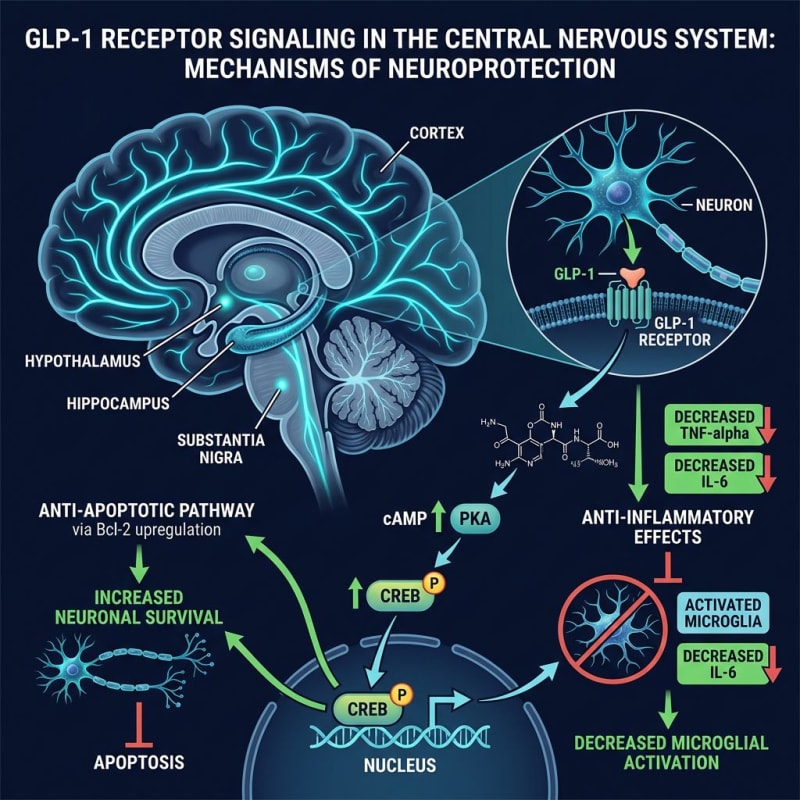
For in-vitro work, establish receptor expression and functional responsiveness in the actual cell preparation rather than assuming that all neuronal, astrocytic, or microglial models behave alike. Cell origin, differentiation state, culture conditions, and receptor-detection methods are important parts of the model description.
Candidate signaling readouts include cAMP, protein kinase A (PKA)-associated responses, CREB phosphorylation, and PI3K/Akt pathway measurements. These should be presented as assay endpoints, not as a guaranteed semaglutide response. Receptor-negative comparators, genetic perturbations, or appropriately characterized pharmacological controls can help test whether an observed response is receptor-dependent.
Inflammatory Readouts
In an in-vitro inflammatory assay, TNF-α, IL-1β, and IL-6 can be measured alongside cell viability and cell number. A lower cytokine signal needs to be distinguished from reduced viable cell abundance or analytical interference. Broad M1/M2 labels should not substitute for a defined marker panel and a description of the cell system. No semaglutide-specific shift in microglial state is asserted here.
Mitochondrial and Cell-Stress Readouts
In cell-based experiments, mitochondrial membrane potential, oxidative-stress measurements, and Bcl-2 or Bax abundance address different aspects of cell state. No single endpoint establishes mitochondrial preservation or neuronal survival. Orthogonal measurements, assay-interference controls, and normalization to viable cell number help make these results interpretable.
Protein Processing and Turnover
In an in-vitro protein-processing model, amyloid-beta abundance, tau phosphorylation, and autophagy-related markers require separate interpretation. A decrease in protein abundance does not by itself demonstrate clearance; altered production, release, or cell number could also affect the measurement. Similarly, a static autophagy marker does not establish autophagic flux. The supplied material is insufficient to attribute specific changes in these endpoints to semaglutide in a defined model.
Distinguishing Evidence Types
| Evidence type | Essential context | Interpretation limit |
|---|---|---|
| In vitro | Cell source, receptor status, assay conditions, and controls | Cell responses do not establish whole-organism exposure or neurological outcomes. |
| Animal | Species, strain, model, tissue, measured endpoint, and experimental controls | Results remain specific to the reported model and cannot establish human effects. |
| Human | Actual study population, study design, tested material, and measured outcomes | Human study findings do not characterize a supplier's nonclinical research material. |
| In silico | Computational method, assumptions, input data, and validation | Predictions do not demonstrate transport, receptor activity, or biological outcomes. |
No human-population or in-silico outcome is summarized here. The original article also asserted outcomes in MPTP-induced rodent models and in ischemic injury models without sufficient verified study details. Those assertions, and claims concerning traumatic brain injury, motor performance, alpha-synuclein, and reward-related behavior, are not carried forward.
Comparators and Receptor Selectivity
Related compounds have different receptor-target profiles. These differences can inform the framing of a receptor-selectivity question, but do not establish equivalent CNS exposure or justify claims of combined effects.
| Compound | Receptor targets |
|---|---|
| Semaglutide | GLP-1 receptor |
| Tirzepatide | GIP and GLP-1 receptors |
| Retatrutide | GIP, GLP-1, and glucagon receptors |
| Cagrilintide | Amylin receptors |
This target summary is not a comparison of potency, brain distribution, or experimental performance. The supplied material does not support recommendations to combine semaglutide with other peptides.
Laboratory Handling and Reproducibility
Follow the applicable safety data sheet, institutional chemical-hygiene procedures, and documented material-specific handling and storage requirements. This article does not supply a hazard classification or validated stability conditions; absence of those details should not be interpreted as absence of hazards.
Record lot identity, available analytical results, storage history, assay timing, and sample handling. Include appropriate matrix and vehicle controls, assess analytical interference, and distinguish technical replicates from independent biological experiments. For animal-derived data, distinguish direct tissue measurements from whole-organism behavioral endpoints and report relevant systemic confounders rather than assigning every change to a direct CNS mechanism.
Supplied References
- Hölscher C. (2022). Protective effects of GLP-1 mimetics in Alzheimer's disease: intracellular signaling and cytoprotective effects. Frontiers in Aging Neuroscience.
- Athelogou M., et al. (2021). Semaglutide, a glucagon-like peptide-1 receptor agonist with protective effects in the central nervous system. CNS Drugs.
- Grieco M., et al. (2019). Glucagon-Like Peptide-1 receptor agonist as a multi-target drug for neurodegenerative diseases. International Journal of Molecular Sciences. — https://doi.org/10.3390/ijms20092230
- Salameh T.S., et al. (2020). Blood-brain barrier transport of glucagon-like peptide-1 receptor agonists. Journal of Cerebral Blood Flow & Metabolism.
- Kim D.S., et al. (2017). GLP-1 receptor activation and neuroprotection in ischemia and neurodegeneration. Journal of Neurochemistry. — https://doi.org/10.1111/jnc.14041
- McLean B.A., et al. (2021). Revisiting the role of GLP-1 receptor signaling in the brain. Molecular Metabolism. — https://doi.org/10.1016/j.molmet.2021.101211
- Zhang L., et al. (2018). Neuroprotective effects of the GLP-1 analog liraglutide and semaglutide in Parkinson's disease models. Scientific Reports. — https://doi.org/10.1038/s41598-018-20984-z
- Drucker D.J. (2021). GLP-1 receptor agonists: non-metabolic actions and clinical implications. Cell Metabolism. — https://doi.org/10.1016/j.cmet.2021.03.001
Limitations
Interpretation requires verification of each study's compound, model, methods, and endpoints. Published findings do not establish safety, effectiveness, approval, or suitability of material sold by Alpha Carbon Labs for human or veterinary use. These materials are intended solely for nonclinical laboratory research.