LL-37 and BPC-157 are distinct peptides discussed in literature concerning tissue responses, inflammation, and extracellular matrix biology. A useful laboratory comparison must distinguish chemical identity, experimental model, measured endpoint, and strength of evidence. The supplied material does not establish a head-to-head comparison of their effects on dermal tissue, macrophage states, or matrix organization.
This article focuses on interpreting that research and defining nonclinical experimental questions. Catalog entries for LL-37 and BPC-157 identify the materials discussed; published observations cannot be attributed to Alpha Carbon Labs materials without direct testing.
Peptide Identity and Scope
LL-37
LL-37 is a 37-amino-acid peptide derived from the human cathelicidin precursor hCAP18. Its identity as a human host-defense peptide does not establish the behavior of a synthetic preparation in a particular assay. Experiments involving purified peptide, endogenous peptide expression, or precursor processing address different questions and should not be treated as interchangeable.
BPC-157
BPC-157 is a synthetic pentadecapeptide: a peptide containing 15 amino acids. Describing the isolated research material as a naturally circulating repair signal, or assigning it a normal physiological function in human skin, is not justified by the information supplied. References to gastric biology also do not establish that a commercial preparation is equivalent to an endogenous material.
For either peptide, interpretation requires the exact test-article identity and its analytical documentation. A shared compound name alone is insufficient to establish comparability between published experiments and a laboratory's material.
A Framework for Studying Dermal Tissue Responses
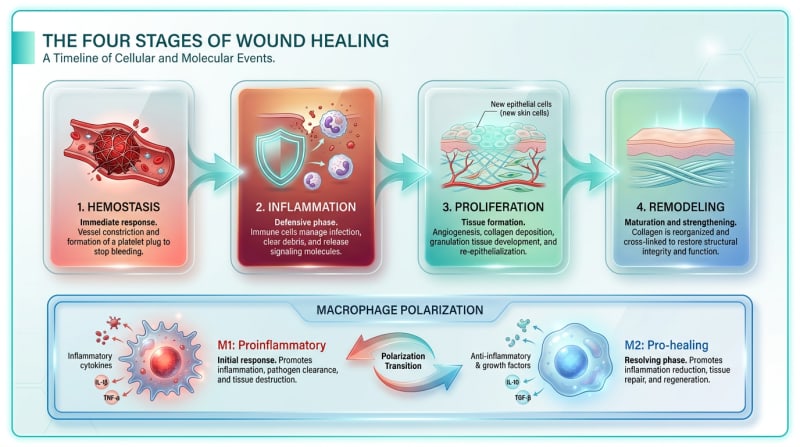
Hemostasis, inflammation, proliferation, and remodeling are conventional, overlapping categories used to describe tissue responses to injury. They are not a fixed timetable or a sequence controlled by one peptide or one macrophage state.
- Hemostasis: clot formation and the development of a provisional matrix.
- Inflammation: immune-cell recruitment, debris processing, and signaling among immune and tissue-resident cells.
- Proliferation: cell proliferation and migration, matrix deposition, and vascular changes.
- Remodeling: changes in matrix composition, organization, turnover, and mechanical properties.
These categories help organize endpoints, but an isolated in-vitro assay reproduces only selected features. For example, a keratinocyte monolayer does not reproduce circulation, immune-cell recruitment, or the architecture of intact dermal tissue.
Macrophage Polarization: Beyond a Binary Switch
The M1/M2 terminology is a simplified framework for macrophage activation, not a complete classification of macrophages in tissue. Macrophage states depend on the cellular source, surrounding signals, and sampling time. An increase in a marker associated with one activation state does not demonstrate a complete transition to another state.
The original claims that LL-37 reliably switches macrophages from M1 to M2, or that BPC-157 has a defined indirect polarization effect, lack sufficiently specified supporting experiments in the supplied material. They therefore cannot serve as established mechanisms or comparative conclusions.
Questions for an In-Vitro Macrophage Study
The following are experimental-design considerations, not findings reported for either peptide:
- Cell source: distinguish primary human cells, primary animal cells, and established cell lines. For primary human cells, document relevant donor characteristics and variability.
- Baseline state: record differentiation conditions, culture medium, and any prior stimulation.
- Readouts: assess multiple activation markers alongside cytokine measurements, viability, and functional endpoints appropriate to the question.
- Time dependence: distinguish an early signaling response from a persistent change in cell state.
- Controls: include matched vehicle and assay-appropriate reference controls, with attention to endotoxin and other potential confounders.
Changes in cytokine release should not automatically be labeled anti-inflammatory, and loss of signal should not be interpreted without evaluating cell viability. Neither a marker profile nor a cytokine measurement alone establishes tissue-level behavior.
Extracellular Matrix: Separate Amount, Structure, and Function
The extracellular matrix includes structural proteins, adhesive proteins, proteoglycans, and other components surrounding cells. In dermal research, matrix deposition and matrix degradation must be distinguished from fiber organization and mechanical properties.
The supplied material does not substantiate claims that either peptide produces a particular collagen architecture or determines a scar outcome. Such claims require defined models, quantitative measurements, and suitable controls rather than a descriptive image alone.
For proposed in-vitro fibroblast or three-dimensional matrix experiments, researchers can distinguish several questions:
- Composition: which matrix components are measured, and how are results normalized?
- Organization: does imaging quantify fiber alignment or distribution rather than total staining alone?
- Turnover: are matrix production and degradation assessed separately?
- Cell contribution: could changes in cell number or viability account for the matrix signal?
- Function: is a mechanical property measured directly, or merely inferred from morphology?
In an in-vitro scratch assay, reduced gap area can reflect migration, proliferation, or both. It should be reported as an assay endpoint, not as proof of intact skin repair. Similarly, endothelial network formation in culture is not equivalent to the formation of functional, perfused vessels in an animal model or a human population.
Comparing LL-37 and BPC-157 Without Ranking Outcomes
| Topic | LL-37 | BPC-157 | Requirement for comparison |
|---|---|---|---|
| Identity | 37-amino-acid cathelicidin-derived peptide. | Synthetic 15-amino-acid peptide. | Document the exact test article and analytical identity. |
| Macrophage response | A consistent M1-to-M2 transition is not established by the supplied material. | A specific polarization mechanism is not established by the supplied material. | Use matched cell sources, controls, sampling times, and multiple readouts. |
| Matrix response | Do not infer matrix organization from cell migration alone. | Do not infer matrix function from collagen staining alone. | Separate composition, organization, turnover, and mechanics. |
| Vascular endpoints | No relative potency ranking is supported here. | No relative potency ranking is supported here. | Distinguish in-vitro endothelial measurements from in-vivo vascular function. |
Different molecular identities or proposed pathways do not establish complementary effects or synergy. The supplied material also does not support extrapolating results from either individual peptide to a multi-component commercial preparation.
Reading the Supplied Literature Critically
The reference list includes titles addressing host-defense peptides, BPC-157, macrophages, and tissue remodeling. A paper title or citation alone does not establish its experimental model, measured outcomes, or relevance to a specific dermal assay. The supplied references are retained unchanged below; bibliographic details and full-text methods should be verified before study-specific findings are used.
No specific human, animal, in-vitro, or in-silico result is asserted here where the supplied information does not identify the underlying model adequately. When extracting evidence, keep these categories separate:
- Human evidence: identify the actual study population and distinguish observational measurements, tissue sampling, and interventional research. Human-derived cells studied outside the body remain in-vitro evidence.
- Animal evidence: identify the species, tissue model, experimental conditions, and measured endpoints. Animal findings do not establish outcomes in humans.
- In-vitro evidence: identify the cell source, culture system, assay conditions, and endpoint. Cellular responses do not establish whole-organism effects.
- In-silico evidence: identify the computational model and validation status. Predictions are not experimental demonstrations.
Reviews provide context but should not be presented as single experiments. Mechanistic claims should be traced to the underlying study and stated alongside its actual model and limitations.
Analytical Documentation and Laboratory Handling
Information about peptide synthesis, quality control, and available Certificates of Analysis can help researchers identify the documentation needed for a test article. These resources are not, by themselves, evidence of a particular lot's identity, purity, biological activity, or suitability for an assay.
Chromatographic purity is method-dependent. An HPLC area percentage is not necessarily peptide mass content and does not independently establish sequence identity, sterility, endotoxin status, or the absence of all impurities. Where available, review identity testing, chromatographic conditions, peptide content, and assay-relevant contaminant measurements separately.
No verified lot-specific analytical specifications, storage conditions, or hazard classification were supplied for this rewrite. Use the material-specific safety data sheet, lot documentation, and institutional laboratory procedures for storage, handling, protective equipment, and disposal. Missing hazard information should not be interpreted as evidence of safety.
Record lot identifiers and relevant handling history when reporting experiments. Analytical characterization and biological assay controls address different sources of uncertainty; neither replaces the other.
References
- Sikiric, P., et al. (2015). The gastrointestinal pharmacology of the gastric juice peptide BPC 157. Current Pharmaceutical Design, 21(23), 3326-3338.
- Gwyer, D., et al. (2019). Gastric pentadecapeptide BPC 157 and its role in healing dermal wounds and promoting angiogenesis. Journal of Investigative Dermatology.
- Tjabringa, G. S., et al. (2005). The antimicrobial peptide LL-37 stimulates angiogenesis and wound healing. Journal of Investigative Dermatology, 125(5), 101-105. — https://pubmed.ncbi.nlm.nih.gov/16098056/
- Kahlenberg, J. M., & Kaplan, M. J. (2013). Little peptide, big effects: the role of LL-37 in inflammation and autoimmune disease. Journal of Immunology, 191(10), 4895-4901. — https://doi.org/10.4049/jimmunol.1302005
- Vukojevic, J., et al. (2018). Pentadecapeptide BPC 157 and the central nervous system. Neural Regeneration Research, 17(3), 482-487. — https://pubmed.ncbi.nlm.nih.gov/34380875/
- Bowdish, C. M. E., et al. (2004). Impact of LL-37 on anti-infective immunity and macrophage function. Journal of Leukocyte Biology, 77(4), 451-459. — https://doi.org/10.1189/jlb.0704407
- Hauk, M., et al. (2020). Macrophage polarization in wound healing: A review of the M1/M2 axis. Tissue Engineering Part B: Reviews.
- Novak, R., et al. (2020). Extracellular matrix remodeling and the regenerative capacity of BPC 157 in tissue injury. Scientific Reports, 10, 15024.
- Mangoni, M. L., et al. (2016). Host defense peptides and skin wound healing. Current Protein & Peptide Science, 17(3), 253-261. — https://pubmed.ncbi.nlm.nih.gov/26601956/
- Wynn, T. A., & Vannella, K. M. (2016). Macrophages in Tissue Repair, Regeneration, and Fibrosis. Immunity, 44(3), 450-462. — https://doi.org/10.1016/j.immuni.2016.02.015